(making sense of complex diagrams)
The medical biochemistry materials make extensive demands on students' ability to visualize structures in three dimensions and to interpret two-dimensional diagrams representing three-dimensional structures. Students must understand the quantitative terminology used to describe shapes and structures. They need to have a well-developed feel for the relative sizes of molecular and cellular structures described or illustrated. Illustrations may combine structures that vary in size by orders of magnitude.

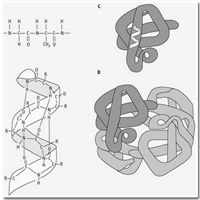
Primary, secondary, tertiary and quaternary structures. (A) The primary structure is composed of a linear sequence of amino acid residues of proteins. (B) The secondary structure indicates the local spatial arrangement of polypeptide backbone yielding an extended α-helical or β-pleated sheet structure as depicted by the ribbon ... (C) the tertiary structure illustrates the three-dimensional conformation of a subunit of the protein; while the quaternary structure (D) indicates the assembly of multiple polypeptide chains into an intact, tetrameric protein. (Baynes & Dominiczak, 2005, p. 18)
In order to make sense of the diagrams students must:
- understand that it comprises several different kinds of representation, ranging from a symbolic representation of a molecule to a semi-representational picture of the 3D structure of the "intact tetrameric protein".
- understand the implicit changes in scale between the diagrams
- understand the meanings of the quantitative terms used in the text.
At the same time they need to navigate the connections between the text in the caption and the diagrams provided.